Signals
- The electric signal is produced during muscle activation, which is observable through the exchange of ions across the muscle membranes and the electrodes attached on the skin.
- EMG signal is acquired through differential amplification.
- sEMG is usually at the range of 5 - 250 Hz (but the cut-off frequency is suggested at 65 - 180 Hz to avoid strong DC at 60 Hz).
Electrodes
- Ag-AgCl electrodes (pre-gelled) are used in over 80% of surface EMG (sEMG) applications due to its low impedance and high stability
- Placement:
Possible noise to sEMG (see more in 2)
- Ambient noise (radiated EMI, specifically 50-60 Hz)
- Transducer noise (different impedence between the skin and electrode sensor)
Other issues
- Consistency in impedance is critical for the reliability of sEMG measurements (think about our form factor)
- Different kind of biosignals - ECG (heart), EEG (brain), EOG (eye) in 3
- Suggestions of electrode placement 3
Fix bug in RXTX library
- The bug in SerialImp.c causing unnecessary delay (~20ms)
- Modify the sleep time from 20000ms to 2000ms, reducing the delay to ~2ms
- Our ADC sampling rate is 256 Hz (i.e., interval ~3.9ms). The 2ms delay fulfills the system requirement.
- Ref:
- Bug issue
- How to compile RxTx: 12
- Install RxTx on Mac
- Setup compiler
- RxTx source code
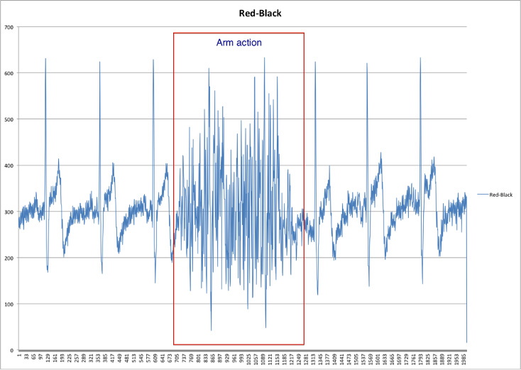
Initial data collection
Keyu implemented a real-time sEMG data retrieval and visualization using Python.